
The SPECIM cameras allow the imaging of any kind of sample without previous preparation in both 400-1000 nm (FX10) and the 900-1700 nm (FX17) using a push-broom system. This allows the system to obtain spectral maps of various samples, enabling chemical analysis of specific regions of the samples. For this reason, hyperspectral imaging systems are ideal for:
- Run non-destructive, non-intrusive, contactless analysis.
- Produce distribution maps of parameters such as moisture, fat or sugar content.
- Classify samples and detect defects, contaminants or foreign bodies.
Acquisition needs very little input from the user. The sample is scanned on a motorised stage under halogen illumination, and the instrument is calibrated against standard references before each measurement, so results can be reproduced from one session to the next.
Spectral range
What is a hyperspectral image?
A conventional camera records three colors per pixel: red, green and blue. A hyperspectral camera records a complete spectrum for every pixel. This image can be seen as a collection of hundreds of photographs of the same scene, each taken at a different wavelength. Each pixel has an “spectral fingerprint” while each of these photographs shows the different regions where sample absorbs or reflects light of a certain wavelength.
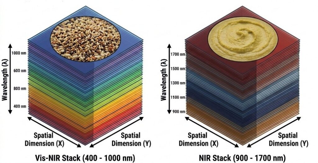
How is the image acquired?
The FX cameras work in push-broom mode: they capture one line of the sample at a time while the sample moves beneath them on a motorised stage. The full image is built up line by line. The scanning is controlled with Specim’s LUMO Scanner software.
Before each measurement, the system is calibrated with two references:
- A white reference, a surface with a known reflectance close to 100%.
- A dark reference, taken with the shutter closed to record the sensor’s own noise.
These references turn the raw signal into relative reflectance. This makes measurements comparable across days, across samples and between the two cameras.
What chemical information can we obtain?
In the near-infrared, light excites overtones and combination bands of molecular vibrations. Each type of chemical bond absorbs in typical regions of the spectrum. Water is a good example:
- It has an absorption band at about 970 nm (second overtone of the O-H stretch), which the FX10 can detect.
- It has a much stronger band at about 1450 nm (first overtone of the O-H stretch), which the FX17 can detect.
By following these bands pixel by pixel, we can see which areas of a fruit, a fillet or a grain hold more moisture.
How do we turn the images into results?
A single hyperspectral image holds hundreds of thousands of strongly correlated spectra. To extract useful information, the spectra are first pre-processed: the background is removed and the spectra are corrected for scattering and baseline effects with SNV, MSC or Savitzky–Golay derivatives. They are then modelled with the machine learning methods our group specialises in:
- Random Forest (RF): an ensemble of decision trees that ranks the importance of each wavelength, which helps in designing simpler multispectral systems.
- Support Vector Machines (SVM): find the best boundary between classes, even with small datasets and non-linear relationships.
- Artificial Neural Networks (ANN): learn complex relationships between the spectrum and the target property, for both classification and regression.
These models work pixel by pixel, so the result is a classification or chemical map of the whole sample. To also use spatial context, Deep Learning models can be applied:
Applications
The system is used mainly in the agri-food sector, for example:
- Quality control and ripeness assessment of fruits and vegetables, including sugar content.
- Detection of bruises, rot and fungal contamination.
- Moisture, fat and protein content in meat, fish, cereals, nuts and baked goods.
- Detection of food fraud and adulteration.
- Sorting of seeds, grains and raw materials.
- Identification of plastics and packaging materials.